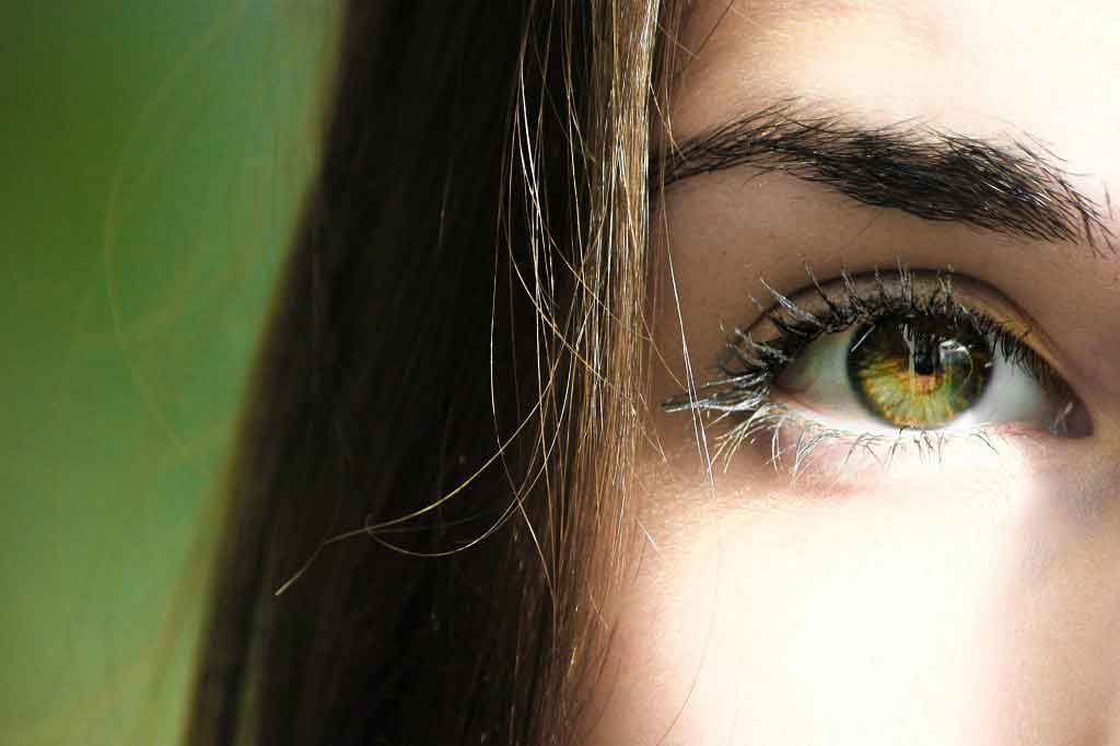
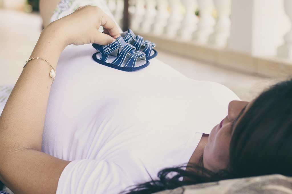
Baby face
Neurology
A baby’s smile “doesn’t just warm a mother’s heart – it gives her a natural high,” reports the Daily Mail. The sight of a smiling infant can trigger the “feel-good”
A baby’s smile “doesn’t just warm a mother’s heart – it gives her a natural high,” reports the Daily Mail . The sight of a smiling infant can trigger the “feel-good” part of the brain, the newspaper says.
The story is based on a small study which profiled the response of 28 mothers to seeing their child’s facial expressions in comparison with an unknown child’s face. Perhaps not surprisingly, the centres associated with pleasure were activated at the sight of smiling babies, and more so if the baby belonged to the mother. The findings may contribute to an understanding of how mothers bond with their babies. However, the practical use of such increased understanding is not currently clear.
Where did the story come from?
Dr Lane Strathearn and colleagues from Baylor College of Medicine in Texas and from University College London carried out this research. The study was funded by the National Institutes of Health, the Baylor Child Health Research Center, the Kane Family Foundation, the National Institute of Neurological Disorders and Stroke, and the National Institute on Drug Abuse. It was published in the peer-reviewed medical journal: Pediatrics .
What kind of scientific study was this?
This was a cross-sectional study of women enrolled during their third trimester into a larger study of mother–infant attachment. In this publication, the researchers explored how particular regions of a mother’s brain (dopamine-associated reward processing areas known to be involved in the pleasure response) are activated in response to pictures of her infant experiencing different emotions.
Women were recruited from a variety of community settings, including prenatal clinics and local church groups, as well as through poster, magazine and internet adverts. The first-time mothers had not given birth to twins, they were all right-handed, non-smoking, were not currently taking psychotropic medications and did not have any contraindications to magnetic resonance imaging (MRI). Demographic information was collected from the eligible women, who also underwent a battery of tests to assess mental health, IQ and the type of relationships that mothers form with other people.
When the infants were seven months old, the researchers videotaped their facial expressions as they responded to different scenarios, including being left along in a room (where they cried) and playing with them using age-appropriate toys (where they smiled). Mothers were not present during this videotaping. The researchers then captured still images of happy, neutral and sad faces of each infant. They also captured facial expressions of a “control” child (i.e. not belonging to any of the women in the study), which was matched to each infant for age, race and sometimes gender. The pictures were captured in a standard way, with the babies wearing a gender-neutral white jumpsuit.
Seven to 17 months after the videotaping, the mothers attended an interview followed by brain scanning, using an MRI. The interview used the Parent Development Interview to prompt mothers to think about their relationship with their infant. Following this, the MRI scan was performed while the women viewed 60 images of an infant’s facial expressions – 30 of her own child and 30 from the matched control. The images, which were randomly presented, contained equal numbers of happy, sad and neutral images. They were presented in a random order. After the scanning session, the images were shown for a second time and the mothers were asked to record what she thought each infant was feeling, as well as her own emotional response.
Although 43 mothers were originally eligible for the study, brain images were available from only 28. Researchers compared these mothers’ brain responses to their own infants with their responses to control infants, and then assessed the effect of the different emotions they had captured.
What were the results of the study?
Overall, regardless of what emotion was being expressed, significantly different areas of the maternal brain were activated by seeing their own infant compared with seeing the control infant. Similarly, there was greater activation in six brain regions (five in the limbic area, one in the midbrain – regions that are involved in emotions, cognition and behaviour) when the mothers’ own happy child was shown compared with an unknown happy child.
With the neutral faces, four of these six areas were significantly more activated with the mother’s own child than the control. With the sad faces, there was no difference between own child and the control in activation in these areas.
Other tests confirmed that the pattern of response in these regions was high activation at happy faces, less activation to neutral faces and none to sad faces. In other brain areas – anterior cingulate, insula and amygdala – sad faces produced widespread activation, and these were more pronounced with the mother’s own child. Not surprisingly, the brain responses correlated with what the mothers reported the infants to be feeling, and their responses were more accurate in the case of their own children.
What interpretations did the researchers draw from these results?
When first-time mothers view their own child’s face, reward-processing regions of the brain are activated. Though it was surprising that there was no difference in response to a mother’s own child crying and an unknown child crying, the researchers conclude that, in this sample of women at least, the results suggest that mothers respond equally to known and unknown children in distress.
What does the NHS Knowledge Service make of this study?
The researchers raise the following point in connection with their results:
- Participating mothers were interviewed and underwent brain scanning when their infants were at different ages. It is possible that the maternal response changes to her child changes over time. Therefore, studying mothers at exactly the same time point after birth may have given different results.
- The researchers say that this study “takes us one step closer to understanding the underlying brain processes and pathways involved in maternal–infant relationships”.
Although these results will feed into other ongoing and future studies that seek to understand the complex nature of the mother-child bond, it is not clear exactly what the practical value of knowing which parts of the brain react to these stimuli might be.
Article Metadata
Date Published: Tue, 22 Aug 2017


 Subscribe
 Ask the doctor
 Rate this article
 Find products