Could new tests use sugar to help detect cancer?
Cancer
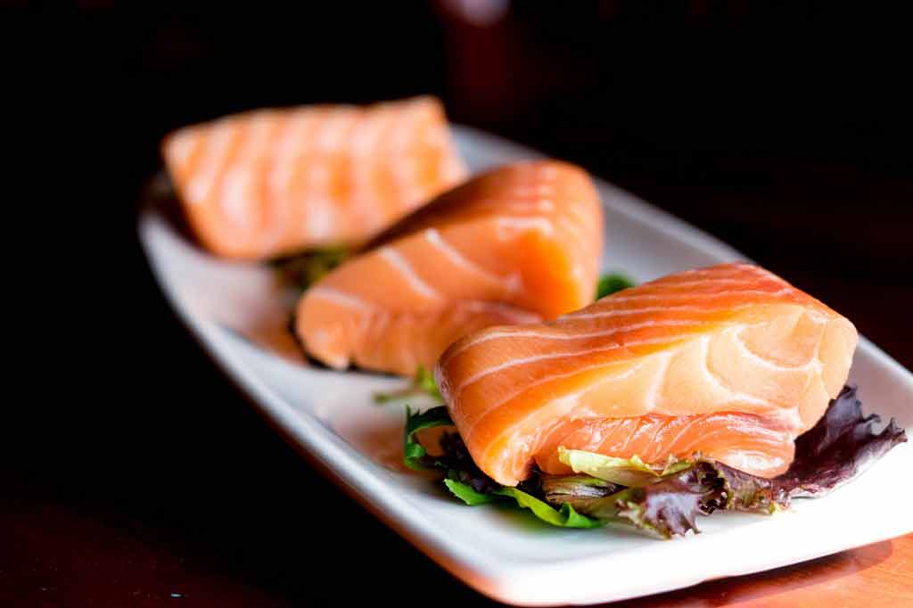
"Chocolate, fizzy drinks and other sugar-laden foods could soon be used to detect cancer," the Mail Online reports. This news is certainly a good way to increase the reader appeal of a very technical study that…
"Chocolate, fizzy drinks and other sugar-laden foods could soon be used to detect cancer," the Mail Online reports.
This news is certainly a good way to increase the reader appeal of a very technical study that looked at whether the way tumours deal with sugar can help in their detection.
Everybody loves chocolate, but the mice involved in this study did not indulge in these sugary treats. Instead, they were given an injection of glucose into their abdominal cavity and then had a new scanning technique called GlucoCEST, which is based on magnetic resonance imaging (MRI). The technique is designed to look for the increased level of glucose intake, which is a hallmark of cancerous tissue.
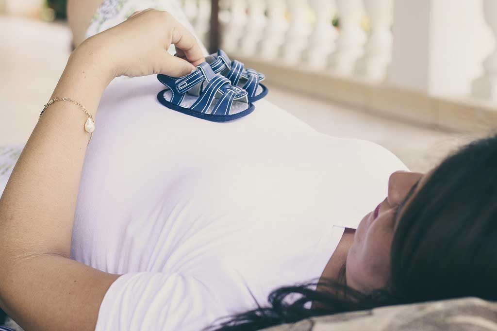
The research showed that the GlucoCEST technique had a similar performance to an established cancer imaging technique called FDG-PET when identifying tumour tissue. This new technique also avoids the need to use radioactively labelled glucose. This means it could be used more frequently and in pregnant women and young children, who are advised to avoid radioactivity where possible.
The Mail Online says that the method "has been trialled on a handful of cancer patients, with early signs of success". This human research was not described in the current publication, so its results are not clear. Further tests in humans are needed to determine whether the technique could be a useful tool in the diagnosis of cancer.
Where did the story come from?
The study was carried out by researchers from University College London (UCL) and other research centres in the UK. It was funded by King's College London and UCL Comprehensive Cancer Imaging Centre, The Institute of Cancer Research Cancer Imaging Centre, Cancer Research UK, the Engineering and Physical Sciences Research Council (EPSRC), the Medical Research Council, the Department of Health, and the British Heart Foundation.
It was published in the peer-reviewed journal Nature Medicine.
The Mail Online's reporting covers the main points of the study. However, the human research reported was not outlined by the scientific paper the story is based on, so the exact details of any ongoing human research and its results are not clear.
What kind of research was this?
This study looked at whether the way tumours deal with sugar can help in their detection. The normal way our cells break down sugar to get energy requires oxygen, but cells can also break down sugar without using oxygen if there is a limited supply. Tumour cells are more reliant on this oxygen-free method of breaking down sugar, and so take up more glucose.
The researchers wanted to find out if they could take advantage of these differences to help them detect tumours in the body using MRI. These differences are already exploited in the detection of metastatic cancer (cancer that spreads from its point of origin to other parts of the body) using a technique called FDG-PET, but this technique uses radioactively labelled glucose. The researchers report that a technique using MRI without radioactivity would be substantially cheaper than FDG-PET.
The researchers used various laboratory and animal experiments, which are appropriate early stage experiments to carry out before moving on to human studies.
What did the research involve?
The researchers called their technique GlucoCEST (glucose chemical exchange saturation transfer). It works by magnetically labelling glucose in the body and measuring changes in the magnetic resonance of water molecules caused by the uptake of this glucose. This is translated into differing brightness levels on the cross-sectional picture of the tissue being scanned.
For their experiments, the researchers used two mouse models of human cancer. The mice had human colorectal (bowel) cancer cells transplanted into their bodies.
Researchers injected glucose into the abdominal cavity of the mice and then used MRI to look at the tumours' glucose uptake. They say the human equivalent of the amount of glucose they used would be 14g, which is roughly the amount of glucose found in half a standard-sized chocolate bar.
The researchers then compared the performance of GlucoCEST and FDG-PET in detecting these tumours. After 24 hours, they used the FDG-PET technique to look at the tumours using radioactively labelled glucose.
What were the basic results?
The researchers found that the GlucoCEST technique could identify the differences between glucose uptake in tumour tissue and normal muscle tissue.
GlucoCEST showed that tumours in one of the mouse models had a lower uptake of glucose than the other mouse model. These were similar findings to the FDG-PET technique. The pattern of glucose uptake within the tumours detected was also similar.
How did the researchers interpret the results?
The researchers concluded that the glucoCEST technique "has potential as a useful and cost-effective method for characterising disease and assessing response to therapy in the clinic".
Conclusion
These early stage results in mice suggest that the glucoCEST technique might be a new way to identify and monitor tumour tissue. It had a similar performance to a technique currently used in cancer imaging called FDG-PET.
The researchers say that glucoCEST is cheaper than FDG-PET, and also has the advantage of not using radioactively labelled glucose. This means it could be used more frequently than FDG-PET without concerns about accumulating radioactive exposure. It would therefore be suitable for people who are more vulnerable to the risks of radiation, such as pregnant women or young children.
However, there are limitations that need to be overcome. For example, the researchers note that the magnetic field strength used in their MRIs was higher than in normal medical MRI machines.
They say that the effect the lower field strength has on their results needs to be tested. If it is not as effective, scientists also need to consider if exposing people to the higher field strength is safe.
The Mail Online suggests that the technique has been trialled in people with cancer, but this is not reported in the scientific paper, so the methods and results of ongoing human research are unclear.
The researchers do mention the human equivalent of the glucose dose given would be about half a chocolate bar. But it is not yet known whether giving glucose by mouth would work best with the new imaging technique. This needs to be tested further, as the mice received glucose from an injection into their abdominal cavity rather than by mouth.
These early results are likely to lead to further investigation of this technique for detecting tumours. Future studies would ideally look at different types of tumour, as well as addressing the issues mentioned above. This research will help determine whether the technique really could be a useful tool in cancer care.
Article Metadata
Date Published: Tue, 15 Aug 2017


 Subscribe
 Ask the doctor
 Rate this article
 Find products